
 Hospital & Clinic Furniture
 Medical Material
 Orthopedics
 Physiotherapy and Rehabilitation
 Beauty Equipment
 Barber and Hair Salon
 Massage and Spa
 Tattoo Equipment
 Dental Equipment
 Health at Home
 Disposables & consumables
 Emergencies
 Fitness and exercise
 Office Supplies
 Laboratory Equipment
  Veterinary Material
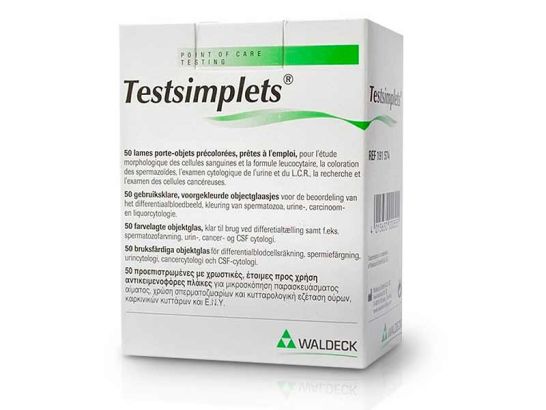
Testsimplets® Prestained Ready-to-Use Slides, box of 50 units.
Manufacturer Information:
Name or company: Pending update
Email: Pending update
Postal address: Pending update
City: Pending update
Country: Pending update
Responsible person information:
Name or company: Pending update
Email: Pending update
Postal address: Pending update
City: Pending update
Country: Pending update
Warnings and product safety information:
The product complies with the General Product Safety Regulation.
Preteñidos slide Testsimplets ® are ready for use, which provide excellent differentiation of the cells contained in body fluids for examination under a microscope and for screening of urine cytology. Testsimplets enables rapid potential differential staining of urine sediment cancer cells, replacing the traditional staining methods to smears and time-consuming Pappenheim.
The easy and clean, and the speed of standard stain colors allow Testsimplets functional use of the consultation and the hospital.
The method of preparation of urine is similar to that used when preparing normal urinary sediment.
1. Centrifuge urine at 3000 rpm for approx. 10 minutes.
2. Carefully decant supernatant.
3. Resuspend the pellet in 3 drops of saline.
4. Place a drop of this suspension in the middle of the colored area of the slide and carefully place the coverslip on top of each included in the kit.
5. After 5-10 minutes has already completed the process of staining.
6. The preparation is now ready for consideration.
The preparation is stable up to 5 hours at room temperature.
Testsimplets is a rapid test in vitro diagnostics for affixing and displaying the differential blood picture under the microscope, being able to dispense with the preparation of a blood frontis
The test consists of a slide equipped with a colored layer. This layer consists of two colors: cresyl violet acetate and new methylene blue. The specific affinity of cellular structures with colors leads to different dyes that can establish a classification of cells. The intensity of color and proportion of mixed colors in the lining of the slide are constant and are standardized, thus obtain reliable staining.
50 slides ready for use, equipped with a layer consisting of 2.1 mg / cm ² of cresyl violet acetate and 1.0 mg / cm ² of new methylene blue.
The slides can be drawn from the case either by hand or with dispenser.
50 coverslips (24 x 36 mm) free from dust
Examine the preparation with an increase of × 100, then identify suspicious cells or cell configurations a × 200 or × 400 increases or oil immersion with an increase of × 1000.
Because vital staining, the cells appear to its natural size, as compared with other methods, details such as the nuclear structure and membranes are clearly visible.
Testsimplets allows accurate differentiation and classification of structures and nuclear membranes, the network of chromatin and nucleoli and cytoplasmic structures.
The urinary cytologic diagnosis is useful in:
all forms of micro-and macroscopic hematuria.
cystitis resistant to treatment.
cistalgia.
dysuria of unknown etiology.
Patient follow-up after urothelial tumor operations.
Early identification and monitoring of bladder cancer.
The determination of the degree of malignancy is made in the same way as with conventional staining techniques.
Ref 191,574
 continue in UK
 Ireland